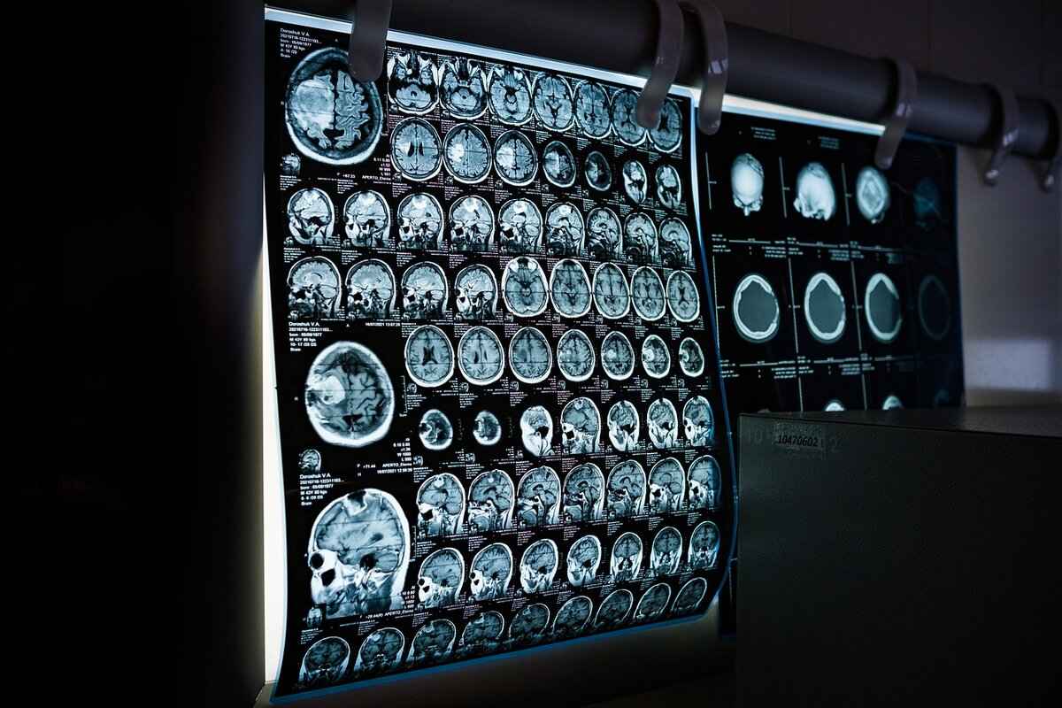
This article explores the process of creating heat maps for CT images, detailing techniques, tools, and applications to enhance medical imaging analysis effectively.
Understanding Heat Maps in Medical Imaging
Heat maps are visual representations that illustrate data density or intensity. In the context of medical imaging, heat maps serve as powerful tools to provide critical insights into the distribution of various parameters within CT images. By converting complex data into an easily interpretable format, heat maps enable healthcare professionals to quickly assess areas of concern and make informed decisions.
Importance of Heat Maps in CT Imaging
Heat maps significantly enhance the interpretation of CT scans by highlighting areas of interest. They play a vital role in diagnosing conditions and guiding treatment decisions through visual data representation. For instance, a heat map can reveal abnormal tissue density, which may indicate the presence of tumors or other anomalies, thus facilitating timely intervention.
Tools Required for Creating Heat Maps
Several software tools and programming languages can be utilized to create heat maps. Understanding the available options is crucial for accurate and efficient mapping of CT images. Popular tools include:
- MATLAB: Known for its powerful data processing capabilities.
- Python: With libraries like Matplotlib and Seaborn, it offers flexibility in customization.
- Specialized medical imaging software: Tailored for healthcare applications.
Step-by-Step Guide to Drawing Heat Maps
Creating a heat map involves several essential steps:
- Data Preparation: Preprocess the CT image data through normalization and filtering.
- Visualization: Select appropriate color gradients to enhance visibility.
- Interpretation: Analyze the heat map data to draw clinical conclusions.
Interpreting Heat Maps in CT Images
Interpreting heat maps requires a solid understanding of the underlying data and its clinical implications. Key features in heat maps, such as hotspots, can indicate areas of concern. Recognizing these features is crucial for making informed medical decisions based on CT images. However, caution is necessary to avoid common pitfalls, such as overemphasizing color intensity, which can lead to misinterpretations.
Applications of Heat Maps in Clinical Practice
Heat maps have diverse applications in clinical settings, from oncology to cardiology. Their ability to visualize complex data makes them invaluable tools for healthcare professionals. For instance, in oncology, heat maps can help visualize tumor metabolism and response to treatment, providing essential insights into patient management and therapeutic effectiveness. In radiology, heat maps enhance the interpretation of CT scans, aiding in the detection of abnormalities and improving diagnostic accuracy.
Future Trends in Heat Mapping for CT Images
The future of heat mapping in CT imaging is promising, with advancements in artificial intelligence (AI) and machine learning poised to revolutionize how heat maps are generated and interpreted. AI can automate the heat map generation process, improving speed and accuracy, which can lead to more efficient workflows in medical imaging. Additionally, emerging visualization techniques, such as augmented reality, are set to enhance the way heat maps are presented, providing immersive experiences for medical professionals.

Understanding Heat Maps in Medical Imaging
Heat maps are powerful visual tools that represent the distribution and intensity of data across various parameters in medical imaging, particularly in CT scans. By employing color gradients, these maps allow healthcare professionals to quickly identify areas of interest and potential abnormalities within the imaging data. The ability to visualize complex data in a simplified manner enhances diagnostic accuracy and aids in clinical decision-making.
In the context of CT imaging, heat maps serve as a bridge between raw data and clinical interpretation. They transform numerical values into a visual format that is easier to analyze, making it possible to spot patterns that may not be immediately apparent from the images alone. For instance, heat maps can reveal regions of heightened metabolic activity, which may indicate the presence of tumors or other pathological conditions.
One of the key advantages of using heat maps in medical imaging is their ability to provide a comprehensive overview of the data. By aggregating information from multiple slices of a CT scan, heat maps can present a three-dimensional perspective of the data, allowing for a more holistic view of the patient’s condition. This capability is particularly useful in complex cases where traditional analysis may fall short.
Furthermore, heat maps can be customized to highlight specific parameters relevant to a particular diagnosis. For example, in oncology, heat maps can illustrate areas of increased blood flow or metabolic activity, which are critical for assessing tumor viability and treatment response. This customization ensures that the heat map serves the specific needs of the clinician, enhancing its effectiveness as a diagnostic tool.
However, the interpretation of heat maps requires careful consideration. Clinicians must be aware of the context in which the data was collected and the implications of the visualized results. Misinterpretations can lead to incorrect diagnoses or treatment plans. Therefore, it is essential for healthcare professionals to be trained in both the creation and interpretation of heat maps to maximize their utility in clinical practice.
In conclusion, heat maps represent a significant advancement in the field of medical imaging. Their ability to visualize complex data makes them invaluable tools for improving diagnostic accuracy and enhancing patient care. As technology continues to evolve, the integration of heat maps into routine clinical practice is likely to expand, offering even greater insights into patient health.

Importance of Heat Maps in CT Imaging
Heat maps are an invaluable tool in the field of medical imaging, particularly when it comes to the interpretation of CT scans. By transforming complex data into a visual format, heat maps enable healthcare professionals to quickly identify areas of interest and potential abnormalities. This visual representation enhances the diagnostic process, making it easier for radiologists and clinicians to make informed decisions regarding patient care.
One of the primary advantages of heat maps is their ability to highlight areas of concern within a CT image. For instance, regions that exhibit abnormal metabolic activity can be easily identified, allowing for more precise diagnoses. This is particularly crucial in oncology, where early detection of tumors can significantly impact treatment outcomes. By visualizing the intensity of signals within the scan, heat maps help to pinpoint hotspots that may indicate malignancies or other critical conditions.
Moreover, heat maps facilitate a more comprehensive analysis of CT images by providing a layered view of the data. Instead of merely examining the grayscale images traditionally associated with CT scans, healthcare providers can observe the distribution of various parameters, such as blood flow or tissue density. This multi-dimensional perspective allows for a deeper understanding of the underlying pathology, ultimately leading to better treatment planning.
In addition to enhancing diagnostic accuracy, heat maps also play a significant role in guiding treatment decisions. For example, in the context of radiation therapy, heat maps can illustrate the response of tumors to treatment over time. By comparing heat maps from different time points, clinicians can assess the effectiveness of therapy and adjust treatment plans accordingly. This dynamic approach to patient care ensures that interventions are tailored to individual needs, improving overall outcomes.
Furthermore, the use of heat maps in CT imaging aligns with the growing trend towards data-driven medicine. As healthcare increasingly relies on quantitative data to inform clinical decisions, heat maps provide a clear and intuitive way to visualize complex datasets. This not only aids in the interpretation of current scans but also contributes to the accumulation of knowledge that can inform future practices and research.
Lastly, it is essential to recognize the educational value of heat maps in the context of medical training. By utilizing these visual tools, medical students and residents can develop a stronger understanding of anatomy and pathology. Heat maps serve as an effective teaching aid, allowing learners to visualize the implications of their findings in a way that traditional images cannot. This enhanced learning experience ultimately prepares the next generation of healthcare professionals to utilize advanced imaging techniques effectively.
In summary, the importance of heat maps in CT imaging cannot be overstated. They not only improve diagnostic accuracy and treatment planning but also contribute to the educational development of medical professionals. As technology continues to advance, the integration of heat maps into clinical practice will likely become even more prevalent, further enhancing the quality of patient care.

Tools Required for Creating Heat Maps
Creating effective heat maps for CT images requires the right tools and technologies. Understanding the various software and programming languages available can significantly enhance the accuracy and efficiency of your mapping process. In this section, we will explore the essential tools required for creating heat maps, focusing on their functionalities and applications in the medical imaging field.
When it comes to creating heat maps, several software options stand out due to their robust features and user-friendly interfaces. Here are some popular choices:
- MATLAB: A powerful tool widely used in engineering and scientific research, MATLAB offers extensive libraries for image processing and visualization. Its built-in functions make it easy to generate heat maps from complex datasets.
- Python: With libraries like Matplotlib and Seaborn, Python is a versatile programming language that provides excellent capabilities for creating custom heat maps. Its flexibility allows for tailored visualizations suitable for specific analytical needs.
- R: Known for its statistical computing capabilities, R is another excellent option for generating heat maps. Packages like ggplot2 enable users to create sophisticated visualizations with ease.
- ImageJ: This open-source image processing program specializes in scientific imaging. It is particularly useful for medical applications, allowing users to analyze and visualize CT images effectively.
- Specialized Medical Imaging Software: Various software solutions are specifically designed for medical imaging, such as OsiriX and 3D Slicer. These tools often come with integrated heat mapping functionalities tailored for healthcare professionals.
In addition to traditional software, programming languages play a crucial role in creating custom heat maps. Here’s how they can be leveraged:
- Python: Beyond its libraries, Python allows for extensive customization. Users can manipulate datasets, apply different statistical methods, and create visually appealing heat maps that cater to specific research questions.
- R: R’s data analysis capabilities are enhanced by its visualization libraries, making it an excellent choice for researchers looking to present their findings through heat maps.
- JavaScript: For web-based applications, JavaScript libraries like D3.js enable the creation of interactive heat maps that can be integrated into web platforms, providing dynamic visualizations for users.
When selecting a tool for heat map creation, consider the following factors:
- Ease of Use: Choose a tool that matches your technical expertise. Some software may have a steeper learning curve than others.
- Specific Features: Identify the features you need, such as data import options, visualization capabilities, and customization levels.
- Integration: Consider how well the tool integrates with other software or systems you are using, especially in clinical settings.
- Cost: While many tools are available for free, others may require a subscription or purchase. Assess your budget accordingly.
By understanding the various tools and programming languages available for heat map creation, professionals can ensure accurate and efficient mapping of CT images. With the right resources, creating insightful visualizations becomes a streamlined process, ultimately enhancing the interpretation of medical data.
Popular Software for Heat Map Creation
Creating heat maps for CT images is a crucial task in medical imaging analysis, and selecting the right software can significantly enhance the process. There are several popular software solutions available that cater to different needs and expertise levels. This section will delve into these tools, highlighting their features, advantages, and how they can streamline the creation of heat maps.
- MATLAB: MATLAB is a powerful tool widely used in academia and industry for numerical computing and data visualization. Its extensive built-in functions allow for the easy generation of heat maps from CT image data. With the Image Processing Toolbox, users can preprocess images, apply filters, and visualize data in a heat map format. The ability to customize visualizations through scripting makes MATLAB a favorite among researchers and professionals.
- Python: Python has gained immense popularity due to its versatility and the vast array of libraries available for data analysis and visualization. Libraries such as Matplotlib and Seaborn are particularly useful for creating heat maps. Matplotlib provides a comprehensive framework for plotting, while Seaborn simplifies the process of creating attractive statistical graphics. These libraries allow users to manipulate data easily, making it possible to generate tailored heat maps that meet specific analytical requirements.
- R: R is another programming language that excels in statistical analysis and data visualization. Packages like ggplot2 and heatmaply facilitate the creation of heat maps with minimal coding effort. R’s focus on statistical modeling makes it an excellent choice for researchers looking to analyze complex datasets derived from CT images.
- Specialized Medical Imaging Software: Several specialized software solutions are designed specifically for medical imaging. Tools like OsiriX and 3D Slicer offer advanced features for visualizing and analyzing medical images. These platforms often include built-in functionalities for generating heat maps, allowing healthcare professionals to visualize data without extensive programming knowledge.
In addition to these software options, many tools offer user-friendly interfaces that facilitate heat map creation without requiring deep technical expertise. This accessibility is vital in clinical settings, where time is of the essence, and healthcare professionals must quickly interpret CT images.
When choosing software for heat map creation, it is essential to consider factors such as the level of customization required, ease of use, and the specific features that align with your analytical needs. By leveraging these popular software solutions, medical professionals can enhance their diagnostic capabilities and improve patient outcomes through more effective data visualization.
Overall, the landscape of software tools available for creating heat maps is diverse and continually evolving. As technology advances, new features and functionalities are likely to emerge, further enhancing the ability to analyze and interpret CT images in clinical practice.
Using Programming Languages for Custom Heat Maps
Programming languages such as Python and R have become essential tools in the realm of data visualization, particularly for creating custom heat maps. These languages offer users the flexibility to manipulate data and tailor visualizations to meet specific analytical needs, making them invaluable in fields like medical imaging, data science, and business intelligence.
- Python: Known for its simplicity and readability, Python has a rich ecosystem of libraries that facilitate the creation of heat maps. Libraries like Matplotlib and Seaborn provide powerful functionalities for data visualization, allowing users to create aesthetically pleasing and informative heat maps with minimal code.
- R: R is a language specifically designed for statistical analysis and data visualization. Packages such as ggplot2 and heatmaply enable users to generate complex heat maps that can include clustering and hierarchical representation of data, which is particularly useful in analyzing large datasets.
When using these programming languages, the process of creating a heat map generally involves several key steps:
1. Data Import: Load the dataset into the programming environment.2. Data Cleaning: Preprocess the data to remove any inconsistencies or outliers.3. Data Transformation: Reshape the data if necessary, ensuring it is in the right format for visualization.4. Heat Map Generation: Use built-in functions or libraries to create the heat map.5. Customization: Adjust color scales, labels, and other visual elements to enhance clarity and interpretability.
One of the primary advantages of using programming languages for heat map creation is the ability to customize visualizations extensively. Users can modify color gradients, adjust the scale of the data, and even incorporate additional layers of information, such as annotations or overlays. This level of customization is crucial in fields like medical imaging, where the ability to highlight specific areas of interest can significantly impact diagnosis and treatment planning.
Moreover, the integration of these programming languages with data analysis frameworks allows for seamless execution of complex analyses. For instance, in Python, one can easily combine data manipulation libraries like Pandas with visualization libraries, facilitating a streamlined workflow from data extraction to visualization.
In addition, the open-source nature of Python and R means that a vast community continuously contributes to their development. This results in a plethora of resources, tutorials, and forums where users can seek help and share knowledge. As a result, individuals and organizations can leverage these tools without incurring high costs, making advanced data visualization accessible to a broader audience.
In conclusion, programming languages like Python and R not only provide the necessary tools to create custom heat maps but also empower users to engage with their data in a meaningful way. The flexibility, customization options, and community support make these languages ideal choices for anyone looking to enhance their data visualization capabilities, particularly in fields that rely heavily on visual data interpretation, such as medical imaging and research.

Step-by-Step Guide to Drawing Heat Maps
Creating a heat map is a meticulous process that involves several essential steps to ensure the final product is both accurate and informative. This guide will walk you through each phase, from the initial data preparation to the final visualization and interpretation of your heat map.
1. Data Preparation for Heat Maps
The first step in drawing a heat map is data preparation. This involves collecting and cleaning the data to ensure its accuracy and relevance. Key actions during this phase include:
- Normalization: Adjusting the data to a common scale without distorting differences in the ranges of values.
- Filtering: Removing noise and irrelevant information that could skew the results.
- Formatting: Ensuring the data is structured properly, often in a matrix format, which is essential for heat map generation.
2. Choosing the Right Visualization Tools
After preparing the data, selecting the appropriate tools for visualization is crucial. Various software options exist, including:
- Python Libraries: Libraries such as Matplotlib and Seaborn are popular for their flexibility and ease of use.
- MATLAB: Known for its powerful data processing capabilities, it is widely used in academic and professional settings.
- Specialized Software: Tools tailored for medical imaging can provide additional functionalities specifically designed for heat map creation.
3. Visualizing Data in Heat Maps
Once the data is prepared and tools are selected, the next step is to visualize the data. This involves:
- Selecting Color Gradients: Choosing a color scheme that effectively represents the data intensity. Warm colors often indicate higher values, while cool colors signify lower values.
- Adjusting Parameters: Fine-tuning settings such as scaling and opacity to enhance the visibility of critical features within the heat map.
4. Interpreting the Heat Map
Interpreting the heat map accurately is essential for deriving meaningful insights. Key considerations include:
- Identifying Hotspots: Recognizing areas with high data density, which may indicate significant findings or anomalies.
- Understanding Context: Analyzing the heat map in relation to the underlying clinical data to draw informed conclusions.
5. Common Pitfalls to Avoid
While creating heat maps, it is important to be aware of potential pitfalls that can lead to misinterpretation:
- Overemphasis on Color Intensity: Relying solely on color intensity can obscure important details; always consider the context of the data.
- Ignoring Data Quality: Poor-quality data can lead to misleading results, so ensure thorough data cleaning and validation.
In summary, creating a heat map is a systematic process that requires careful attention to detail at each stage. By following these steps—data preparation, tool selection, visualization, interpretation, and avoiding common pitfalls—you can produce effective heat maps that provide valuable insights in various fields, particularly in medical imaging.
Data Preparation for Heat Maps
Creating accurate and informative heat maps from CT images is a multi-step process that begins with data preparation. This phase is crucial, as the quality and format of the data directly influence the effectiveness of the heat map. Proper preparation includes several key steps: normalization, filtering, and conversion to the appropriate format.
- Normalization: This process adjusts the data to a common scale, ensuring that variations in intensity do not skew the results. Normalization can be performed using various methods, such as min-max scaling or z-score normalization. These techniques help in maintaining consistency across different datasets, allowing for a more accurate representation of the data’s underlying patterns.
- Filtering: Filtering is essential for removing noise and artifacts from the CT images. Techniques such as Gaussian filtering or median filtering can be employed to enhance the quality of the images. By reducing noise, the heat map can more accurately reflect the areas of interest, making it easier to identify significant features.
- Data Formatting: Ensuring that the data is in the correct format is vital for analysis. Most heat map generation tools require data in specific structures, such as matrices or arrays. It is essential to convert the CT images into these formats, often involving the extraction of pixel intensity values and their arrangement into a suitable data structure.
Additionally, metadata associated with the CT images, such as patient demographics and scan parameters, can enhance the context of the heat map. Including this information allows for a more comprehensive analysis, enabling healthcare professionals to draw more informed conclusions from the visualized data.
Another important aspect of data preparation is the selection of regions of interest (ROIs). Identifying ROIs allows analysts to focus on specific areas within the CT images that may indicate abnormality or require further investigation. By concentrating on these areas, the heat map can provide clearer insights into potential health issues.
In summary, the data preparation stage is foundational for creating effective heat maps from CT images. By implementing normalization, filtering, and proper formatting, along with considering metadata and ROIs, analysts can ensure that the subsequent visualization is both accurate and informative. This meticulous approach not only enhances the quality of the heat map but also significantly contributes to improved diagnostic capabilities in clinical practice.
Visualizing Data in Heat Maps
Once the data is prepared, the next crucial step in the heat map creation process is visualization. This phase is essential as it transforms raw data into a visually compelling format that conveys complex information at a glance. Effective visualization allows for the identification of patterns, trends, and anomalies within CT images, which are critical for accurate medical assessments.
To achieve optimal visualization, one must carefully select color gradients that not only enhance aesthetic appeal but also improve the interpretability of the data. Color gradients can significantly influence how data is perceived. For instance, a gradient that transitions from cool colors (like blue) to warm colors (like red) can effectively indicate low to high intensity areas. This method allows healthcare professionals to quickly identify regions of interest, such as tumors or areas of inflammation.
Moreover, adjusting various parameters during the visualization process can further enhance the clarity of significant features. Parameters such as contrast, brightness, and opacity can be manipulated to highlight specific areas of the heat map. For example, increasing the contrast can make subtle differences in data intensity more pronounced, which is particularly useful when analyzing dense medical images.
In addition to color gradients and parameter adjustments, incorporating interactive elements into heat maps can significantly improve user engagement and understanding. Interactive heat maps allow users to hover over specific areas to obtain detailed information, such as exact data values or additional contextual insights. This interactivity not only aids in better data comprehension but also facilitates a more thorough analysis of the CT images.
Furthermore, employing techniques such as normalization can ensure that the data visualized is comparable across different images or datasets. Normalization adjusts the data scale, allowing for consistent representation regardless of variations in the original data. This step is particularly important in medical imaging, where discrepancies in imaging modalities or patient conditions can affect raw data readings.
Lastly, it is essential to consider the target audience when designing heat maps. Different stakeholders, such as radiologists, oncologists, and patients, may require distinct levels of detail and types of information. Customizing the visualization approach to meet the specific needs of these groups can enhance the effectiveness of communication and decision-making in clinical settings.
In summary, the visualization of data in heat maps is a multifaceted process that involves careful selection of color gradients, parameter adjustments, and the incorporation of interactive elements. By focusing on these aspects, healthcare professionals can create heat maps that not only look appealing but also serve as powerful tools for enhancing the analysis of CT images, ultimately leading to better patient outcomes.

Interpreting Heat Maps in CT Images
Interpreting heat maps is a crucial aspect of analyzing CT images, as it provides an intuitive way to visualize complex data. To effectively interpret these maps, one must possess a solid understanding of the underlying data and its clinical implications. Heat maps serve as powerful tools that can significantly impact diagnosis and treatment planning.
Heat maps represent variations in data intensity, allowing healthcare professionals to quickly identify areas of interest within CT images. However, accurate interpretation goes beyond merely observing color gradients; it requires a comprehensive understanding of the anatomical and pathological contexts of the data presented. For instance, a region of high intensity may indicate a tumor or an area of inflammation, but without proper clinical correlation, these findings could lead to misdiagnosis.
One of the primary challenges in interpreting heat maps is the potential for misinterpretation. Factors such as the chosen color palette, scale, and the specific parameters analyzed can all influence the visual representation of data. Thus, it is essential to consider these elements critically. For example, while a bright red spot may seem alarming, it is important to assess the surrounding data and clinical history to draw accurate conclusions.
Moreover, understanding the significance of different features within a heat map is vital. Hotspots, which indicate areas of increased activity or abnormality, must be evaluated in conjunction with other imaging modalities and clinical findings. This holistic approach ensures that healthcare providers make informed decisions based on comprehensive data analysis.
Another important aspect of interpreting heat maps is recognizing common pitfalls. Overemphasis on color intensity can lead to incorrect conclusions, as not all bright areas necessarily indicate critical issues. It is crucial to maintain a balanced perspective and consider the entire context of the image, including patient history and other diagnostic tests.
In clinical practice, the ability to interpret heat maps accurately can enhance patient outcomes. For instance, in oncology, heat maps can reveal tumor metabolism and response to treatment, guiding oncologists in tailoring therapeutic strategies. Similarly, in radiology, heat maps can aid in detecting abnormalities, improving the overall diagnostic accuracy of CT scans.
In conclusion, interpreting heat maps in CT images is a multifaceted process that requires a blend of technical knowledge and clinical insight. By understanding the underlying data, recognizing key features, and avoiding common pitfalls, healthcare professionals can leverage heat maps to enhance their diagnostic capabilities and ultimately improve patient care.
Identifying Key Features in Heat Maps
Heat maps are an essential tool in the analysis of CT images, providing a visual representation of data that can reveal significant insights into patient conditions. Among the various elements that can be identified within heat maps, hotspots are particularly noteworthy. These hotspots often indicate areas of concern, such as the presence of tumors, inflammation, or other pathological changes. Understanding how to identify these key features is crucial for healthcare professionals aiming to make informed medical decisions.
- Definition of Hotspots: Hotspots are regions within a heat map that exhibit a higher concentration of data points compared to surrounding areas. In the context of CT imaging, these areas can signify abnormal tissue behavior or disease progression.
- Color Gradients: The use of color gradients in heat maps plays a vital role in identifying hotspots. Typically, warmer colors such as red or yellow signify higher intensity or density, while cooler colors like blue or green indicate lower values. Recognizing these color patterns helps clinicians quickly pinpoint areas that require further examination.
- Spatial Distribution: Analyzing the spatial distribution of hotspots can provide insights into the nature of the underlying condition. For instance, clustered hotspots may suggest a localized disease process, while dispersed hotspots could indicate systemic issues.
In addition to hotspots, there are other significant features that can be identified in heat maps:
- Trends Over Time: By comparing heat maps generated at different time points, clinicians can observe changes in hotspot intensity or distribution. This temporal analysis is crucial for monitoring disease progression or response to treatment.
- Comparative Analysis: Heat maps allow for the comparison of multiple CT images from different patients or different scans of the same patient. This comparison can reveal variations in disease presentation and help in tailoring individualized treatment plans.
When interpreting heat maps, it is essential to consider the context of the clinical scenario. For example, a hotspot in a lung CT scan may indicate a potential malignancy, but it could also represent a benign condition, such as an infection. Therefore, corroborating heat map findings with clinical history and other diagnostic tests is vital for accurate interpretation.
Moreover, a thorough understanding of the limitations of heat maps is essential. Misinterpretation can occur if the clinician overemphasizes color intensity without considering the overall clinical picture. Awareness of these potential pitfalls ensures that healthcare professionals utilize heat maps effectively, enhancing their diagnostic capabilities.
In summary, identifying key features in heat maps, particularly hotspots, is integral to improving the accuracy of medical diagnoses based on CT images. By leveraging the visual insights provided by heat maps, clinicians can make more informed decisions regarding patient management and treatment strategies.
Common Pitfalls in Heat Map Interpretation
Heat maps are powerful tools in medical imaging, particularly in the analysis of CT scans. However, misinterpretation of these visual representations can lead to incorrect conclusions and potentially misguided clinical decisions. It is essential to recognize and understand the common pitfalls associated with heat map interpretation to ensure accurate analysis and effective patient care.
One significant issue in heat map interpretation is the overemphasis on color intensity. While color gradients are designed to highlight areas of varying data density, relying solely on these colors can mislead analysts. For instance, a bright red area may suggest a high intensity of a particular parameter, but without understanding the context of the data, it could represent a normal variation rather than a pathological condition. Therefore, it is crucial to combine visual cues with a solid understanding of the underlying data.
Another common pitfall is the neglect of spatial context. Heat maps often display data in a two-dimensional format, which can obscure important spatial relationships. For example, a hotspot in a heat map might be interpreted as a significant area of concern, but if it is located in a region that is not clinically relevant, it may not warrant further investigation. Analysts should always consider the anatomical and functional significance of the areas highlighted in the heat map.
Data normalization is also a critical factor in heat map interpretation. If the data used to create the heat map has not been properly normalized, it can lead to skewed results. Analysts must ensure that the data is standardized to allow for accurate comparisons across different regions of the CT scan. Failure to do so can result in misleading interpretations and erroneous conclusions.
Additionally, analysts should be cautious of confirmation bias when interpreting heat maps. This cognitive bias occurs when individuals favor information that confirms their pre-existing beliefs or hypotheses. In the context of heat maps, this could mean overlooking areas that do not support a clinician’s initial diagnosis while overvaluing those that do. To mitigate this risk, it is essential to approach heat map analysis with an open mind and consider all data points objectively.
Finally, the lack of interdisciplinary collaboration can lead to misinterpretation of heat maps. Medical imaging is a multidisciplinary field that involves radiologists, oncologists, and other specialists. Each professional brings unique insights to the table. When interpreting heat maps, it is beneficial to engage in discussions with colleagues from different specialties to gain a more comprehensive understanding of the data and its implications.
In summary, while heat maps are invaluable in the analysis of CT images, awareness of common pitfalls is essential for accurate interpretation. By recognizing the potential for misinterpretation due to overemphasis on color intensity, neglect of spatial context, improper data normalization, confirmation bias, and lack of interdisciplinary collaboration, healthcare professionals can enhance their analytical skills and improve patient outcomes.

Applications of Heat Maps in Clinical Practice
Heat maps have emerged as powerful tools in clinical practice, offering a visual representation of complex data that aids healthcare professionals in making informed decisions. Their versatility spans across various medical fields, enhancing diagnostic accuracy and treatment evaluation. Below, we explore the diverse applications of heat maps in clinical settings, focusing on their contributions to oncology, cardiology, and radiology.
- Heat Maps in Oncology: In oncology, heat maps are instrumental in visualizing tumor characteristics, such as metabolic activity and response to therapies. By mapping the intensity of metabolic processes, oncologists can assess the effectiveness of treatments over time. This visual approach facilitates the identification of viable tumor regions and helps to tailor personalized treatment plans based on individual patient responses.
- Heat Maps in Cardiology: In cardiology, heat maps play a crucial role in analyzing cardiac imaging data. They can illustrate areas of ischemia or infarction, allowing cardiologists to pinpoint regions requiring intervention. By representing blood flow dynamics and myocardial perfusion, heat maps assist in evaluating the severity of heart conditions and guide therapeutic strategies.
- Heat Maps in Radiology: Radiologists utilize heat maps to enhance the interpretation of CT scans and MRIs. By highlighting abnormal areas, heat maps improve the diagnostic process, enabling radiologists to detect subtle changes that may indicate disease progression. This visual tool not only enhances accuracy but also aids in effective communication with referring physicians regarding patient management.
- Heat Maps in Neurology: In neurology, heat maps can illustrate brain activity patterns during various tasks or resting states. This application is particularly valuable in understanding neurological disorders, such as epilepsy or Alzheimer’s disease. By visualizing brain function, neurologists can better assess cognitive impairments and devise appropriate treatment plans.
- Heat Maps in Pathology: Pathologists employ heat maps to analyze tissue samples and identify areas of interest, such as tumor margins or inflammatory responses. This application enhances the accuracy of diagnoses and provides insights into the biological behavior of diseases, supporting more effective treatment decisions.
In summary, the applications of heat maps in clinical practice are vast and impactful. By transforming complex data into easily interpretable visual formats, heat maps empower healthcare professionals to enhance diagnostic accuracy, tailor treatment approaches, and ultimately improve patient outcomes. As technology advances, the integration of heat mapping into routine clinical workflows is likely to expand, further solidifying their role as indispensable tools in modern medicine.
Heat Maps in Oncology
In the field of oncology, the utilization of heat maps represents a significant advancement in the visualization of complex data related to tumor behavior and treatment responses. These visual tools allow oncologists to interpret intricate patterns of tumor metabolism and assess how patients are reacting to various therapies. By translating data into a visual format, heat maps facilitate a more intuitive understanding of the underlying biological processes that govern tumor dynamics.
Understanding Tumor Metabolism through Heat Maps
Tumor metabolism is a critical area of study in oncology, as it provides insight into how cancer cells derive energy and grow. Heat maps can illustrate metabolic activity across different regions of a tumor, highlighting areas with elevated or reduced metabolic rates. This visualization helps clinicians identify regions that may be more aggressive or resistant to treatment. For instance, by employing heat maps, oncologists can pinpoint hotspots of high metabolic activity that may correspond to tumor aggressiveness, thereby tailoring treatment strategies more effectively.
Evaluating Treatment Responses
Another vital application of heat maps in oncology is their role in evaluating treatment responses. By comparing heat maps generated before and after treatment, oncologists can visually assess changes in tumor metabolism. This comparison is essential for determining the efficacy of therapies, as it allows for the identification of responding and non-responding tumor regions. Such insights are crucial for making informed decisions about continuing, modifying, or switching treatment regimens.
Enhancing Patient Management
The integration of heat maps into clinical practice enhances patient management significantly. By providing a clear visual representation of tumor behavior, oncologists can engage in more informed discussions with patients about their conditions and treatment options. Furthermore, heat maps can serve as valuable tools for multidisciplinary teams, enabling better collaboration and shared decision-making regarding treatment plans.
Future Directions in Heat Mapping for Oncology
As technology continues to evolve, the future of heat mapping in oncology looks promising. The integration of artificial intelligence and machine learning algorithms is expected to enhance the accuracy and efficiency of heat map generation. These advancements could lead to more personalized treatment approaches, as AI can analyze vast amounts of data to identify patterns that may not be readily apparent to human observers.
Conclusion
In summary, heat maps are revolutionizing the field of oncology by providing critical insights into tumor metabolism and treatment responses. Their ability to visualize complex data enhances patient management and fosters more informed clinical decision-making. As the technology advances, the potential applications of heat maps in oncology are likely to expand, offering even greater benefits for patient care and treatment outcomes.
Heat Maps in Radiology
Radiology has significantly evolved with technological advancements, particularly in the realm of imaging techniques. One of the most promising developments is the use of heat maps, which serve as powerful tools for enhancing the interpretation of radiological images, particularly CT scans. These visual aids provide radiologists with a more nuanced understanding of the data, ultimately improving diagnostic accuracy.
What are Heat Maps?
Heat maps are graphical representations that utilize color gradients to depict data density or intensity over a specific area. In the context of radiology, they highlight regions of interest within CT scans, allowing for a more focused analysis. By transforming complex data into visual formats, heat maps help radiologists quickly identify abnormalities that may not be immediately evident in standard imaging.
How Do Heat Maps Enhance Diagnostic Accuracy?
The integration of heat maps into radiological practices enhances diagnostic accuracy in several ways:
- Highlighting Abnormalities: Heat maps can effectively pinpoint areas with abnormal metabolic activity, such as tumors or lesions, making it easier for radiologists to identify potential health issues.
- Improving Workflow: By providing a visual representation of data, heat maps streamline the interpretation process, allowing radiologists to make quicker and more informed decisions.
- Facilitating Communication: Heat maps serve as a common language between radiologists and other healthcare professionals, enabling clearer discussions about findings and treatment options.
Applications of Heat Maps in Radiology
Heat maps have a wide range of applications in radiology. They are particularly beneficial in oncology, where they can visualize tumor behavior and treatment response. For instance, heat maps can illustrate areas of increased metabolic activity in tumors, which can be crucial for assessing the effectiveness of chemotherapy or radiation therapy.
In cardiology, heat maps can highlight regions of the heart that may be experiencing reduced blood flow, aiding in the diagnosis of conditions such as ischemic heart disease. Furthermore, heat maps can be used in the analysis of brain scans to identify areas of abnormal activity associated with neurological disorders.
Challenges and Considerations
Despite their advantages, the use of heat maps in radiology is not without challenges. Radiologists must be trained to interpret these visualizations accurately, as misinterpretation can lead to incorrect diagnoses. Additionally, the quality of the underlying data significantly impacts the reliability of the heat maps generated. Therefore, ensuring high-quality imaging and proper data preprocessing is essential for effective heat map utilization.
The Future of Heat Maps in Radiology
The future of heat maps in radiology looks promising, particularly with the integration of artificial intelligence (AI) and machine learning technologies. These advancements can automate the process of generating heat maps, allowing for real-time analysis and enhancing the overall efficiency of radiological practices. As these technologies continue to evolve, the potential for heat maps to revolutionize diagnostic processes in radiology becomes increasingly apparent.
In summary, heat maps are transforming the landscape of radiology by enhancing the interpretation of CT scans and improving diagnostic accuracy. Their ability to visualize complex data in an intuitive manner makes them invaluable tools for radiologists, ultimately benefiting patient care and treatment outcomes.

Future Trends in Heat Mapping for CT Images
The landscape of medical imaging is rapidly evolving, particularly with the integration of advanced technologies such as artificial intelligence (AI) and machine learning (ML). These innovations are set to transform the field of heat mapping in CT imaging, enhancing both the generation and interpretation of heat maps. This article delves into the promising future trends that are shaping the way heat maps are utilized in CT imaging.
Integration of AI in Heat Map Generation
One of the most significant advancements in heat mapping for CT images is the integration of artificial intelligence. AI algorithms can analyze vast amounts of imaging data quickly and accurately, automating the heat map generation process. This not only enhances the speed of analysis but also improves the precision of the resulting heat maps. By leveraging deep learning techniques, AI can identify patterns and anomalies within CT images that may not be immediately evident to the human eye. Furthermore, AI-driven tools can adapt and learn from new data, continuously improving their accuracy and effectiveness over time.
Moreover, AI can assist in data normalization and preprocessing, ensuring that the input data for heat maps is of high quality. This is crucial as the accuracy of heat maps heavily depends on the quality of the underlying data. By automating these processes, healthcare professionals can focus more on interpreting results and making informed decisions, rather than spending time on data preparation.
Advancements in Visualization Techniques
Emerging visualization techniques are also set to revolutionize how heat maps are presented and interpreted. One notable trend is the use of augmented reality (AR) and virtual reality (VR)
Additionally, advancements in color mapping and gradient techniques are improving the clarity of heat maps. Enhanced color palettes can help highlight critical areas more effectively, ensuring that healthcare professionals can quickly identify regions that require further investigation. These improvements not only aid in diagnosis but also enhance communication among medical teams, as visually impactful heat maps can be more easily shared and discussed.
Personalized Medicine through Heat Mapping
As the field of personalized medicine continues to grow, heat mapping will play a crucial role in tailoring treatments to individual patients. By analyzing heat maps in conjunction with patient-specific data, clinicians can gain insights into how a patient’s unique biology responds to various treatments. This level of customization can lead to more effective treatment plans and improved patient outcomes.
Furthermore, heat maps can be instrumental in monitoring treatment efficacy over time. For example, in oncology, heat maps can illustrate changes in tumor metabolism in response to therapy, providing valuable feedback on treatment effectiveness. This dynamic approach to patient care aligns with the broader trend towards personalized healthcare, where treatments are increasingly tailored to the individual rather than a one-size-fits-all model.
Collaborative Tools and Cloud Computing
The future of heat mapping in CT imaging is also being shaped by collaborative tools and cloud computing. With the ability to store and share large datasets in the cloud, healthcare professionals can collaborate more efficiently across different institutions and specialties. This collaborative approach enables the sharing of insights, techniques, and best practices, ultimately enhancing the quality of heat mapping and medical imaging as a whole.
Cloud-based platforms can also facilitate real-time analysis and visualization of heat maps, allowing for immediate feedback and decision-making. This is particularly beneficial in emergency situations where timely interventions can significantly impact patient outcomes. The integration of collaborative tools into heat mapping workflows represents a significant shift towards more connected and efficient healthcare delivery systems.
As we move forward, the future of heat mapping in CT imaging appears promising, with AI, advanced visualization techniques, personalized medicine, and collaborative tools paving the way for more effective and efficient medical imaging practices. These trends not only enhance the capabilities of healthcare professionals but also improve patient care and outcomes in an increasingly complex medical landscape.
Integration of AI in Heat Map Generation
Artificial Intelligence (AI) is transforming the landscape of medical imaging, particularly in the generation of heat maps for CT images. By automating the heat map creation process, AI enhances both the **speed** and **accuracy** of these critical visual tools, which ultimately leads to more efficient workflows in medical imaging.
One of the significant advantages of integrating AI in heat map generation is its ability to analyze vast datasets quickly. Traditional methods of creating heat maps can be time-consuming and prone to human error. However, AI algorithms can process large volumes of imaging data in a fraction of the time, allowing healthcare professionals to focus on diagnosis and treatment rather than data preparation. This efficiency is especially crucial in emergency situations where timely decisions can significantly impact patient outcomes.
Moreover, AI-driven heat map generation improves **accuracy** by minimizing subjective bias. Conventional methods often rely on the expertise and interpretation of radiologists, which can vary. AI systems, trained on extensive datasets, can identify patterns and anomalies with a level of precision that surpasses human capabilities. This objectivity is essential in clinical settings, as it helps to ensure that critical areas of concern are not overlooked.
In addition to speed and accuracy, the integration of AI facilitates the continuous improvement of heat map generation techniques. Machine learning algorithms can learn from previous cases, adapting and refining their analysis methods over time. This adaptability means that as more data becomes available, the quality of heat maps will continue to improve, providing even more valuable insights for medical professionals.
Furthermore, AI can enhance the visualization of heat maps by incorporating advanced graphical techniques. For instance, augmented reality (AR) and virtual reality (VR) can be integrated with AI-generated heat maps, allowing healthcare providers to interact with the data in an immersive environment. This innovative approach not only aids in better understanding complex data but also enhances collaborative decision-making among medical teams.
The applications of AI in heat map generation extend beyond mere visualization. By integrating predictive analytics, AI can help forecast potential health risks by identifying trends and patterns in heat maps. For example, in oncology, AI can analyze tumor metabolism and response to treatments, providing critical insights that can guide therapeutic decisions.
Despite these advancements, it is essential to address the challenges associated with AI integration. Issues such as data privacy, algorithm transparency, and the need for regulatory frameworks must be carefully considered. Ensuring that AI systems are developed responsibly and ethically will be crucial for their acceptance in clinical practice.
In summary, the integration of AI in heat map generation represents a significant leap forward in medical imaging. By improving speed, accuracy, and visualization techniques, AI enhances the diagnostic capabilities of healthcare professionals. As technology continues to evolve, the potential for AI to revolutionize heat mapping and other aspects of medical imaging is immense, paving the way for improved patient care and outcomes.
Advancements in Visualization Techniques
The field of medical imaging is undergoing a significant transformation due to the rapid advancements in visualization techniques. Among these, **augmented reality (AR)** stands out as a groundbreaking method that is reshaping how medical professionals interact with complex imaging data. By merging digital information with the real world, augmented reality provides a unique platform for enhancing the interpretation of heat maps derived from CT images.
- Immersive Experiences: Augmented reality allows healthcare professionals to visualize heat maps in a three-dimensional space, enabling them to see the data in real-time and from various angles. This immersive experience can lead to a better understanding of the anatomical structures and pathological conditions represented in the images.
- Enhanced Data Interaction: With AR, users can manipulate heat maps using gestures or touch, making it easier to focus on specific areas of interest. This interactivity facilitates a deeper analysis of the data, allowing for more informed clinical decisions.
- Integration with Other Modalities: Augmented reality can be integrated with other imaging modalities, such as MRI or PET scans, providing a comprehensive view of a patient’s condition. This integration enhances the diagnostic capabilities of healthcare providers, leading to improved patient outcomes.
Furthermore, the incorporation of **machine learning algorithms** into visualization techniques is revolutionizing how heat maps are generated and interpreted. Machine learning can analyze vast amounts of imaging data, identifying patterns that might not be visible to the naked eye. This capability allows for the creation of more accurate and informative heat maps, which can significantly aid in diagnosis and treatment planning.
For example, in oncology, machine learning can help identify tumor hotspots by analyzing metabolic activity. This information can be visualized in a heat map, guiding oncologists in making treatment decisions based on the most active areas of a tumor.
Additionally, advancements in **virtual reality (VR)** are also contributing to the evolution of visualization techniques. By immersing medical professionals in a virtual environment, VR allows for the exploration of complex anatomical structures and the visualization of heat maps in a way that traditional methods cannot achieve.
- Training and Education: VR can be used as a training tool for medical students and professionals, providing them with hands-on experience in interpreting heat maps and understanding their clinical implications.
- Collaboration: Virtual reality environments can facilitate collaboration between healthcare providers, allowing multiple users to view and interact with the same heat map simultaneously, regardless of their physical location.
As these technologies continue to evolve, the future of heat mapping in medical imaging looks promising. The integration of augmented and virtual reality with advanced machine learning techniques is set to enhance not only the visualization of CT images but also the overall quality of patient care. By providing healthcare professionals with more powerful tools for data interpretation, these advancements will ultimately lead to improved diagnostic accuracy and better treatment outcomes.
In summary, the advancements in visualization techniques, particularly through augmented reality and machine learning, are transforming the landscape of medical imaging. These technologies offer immersive experiences, enhance data interaction, and improve diagnostic capabilities, paving the way for a new era in healthcare.
Frequently Asked Questions
- What is a heat map in medical imaging?
A heat map is a visual representation that displays data density or intensity within medical images, particularly CT scans. It helps in identifying areas of interest by highlighting specific features and patterns, making it easier for healthcare professionals to analyze the data.
- Why are heat maps important for CT imaging?
Heat maps enhance the interpretation of CT scans by providing a clear visual representation of critical areas. They play a vital role in diagnosing medical conditions and guiding treatment decisions, as they can reveal underlying issues that may not be immediately apparent in standard imaging.
- What tools can I use to create heat maps?
There are several tools available for creating heat maps, including software like MATLAB, Python with libraries such as Matplotlib and Seaborn, and specialized medical imaging software. These tools allow for accurate and efficient mapping of CT images, catering to various analytical needs.
- How do I interpret the data presented in a heat map?
Interpreting heat maps involves understanding the underlying data and recognizing key features, such as hotspots, which indicate areas of concern. It’s crucial to be aware of common pitfalls, like overemphasizing color intensity, to ensure accurate analysis and informed medical decisions.
- What are the applications of heat maps in clinical practice?
Heat maps have numerous applications in clinical settings, including oncology and radiology. They help visualize tumor metabolism, monitor treatment responses, and enhance the interpretation of CT scans, ultimately improving diagnostic accuracy and patient management.


